FragLite: cyclin K
Once we can see where a partner binds, we can build tools to block it. Cyclin K helps control the transcription of long, damage-response genes through cyclin-dependent kinases 12 and 13 (CDK12/13). From its FragLite map we made both precise mutants and, using protein-design AI, small custom-built binders that grip cyclin K tightly — a new toolkit for dissecting its interactions.
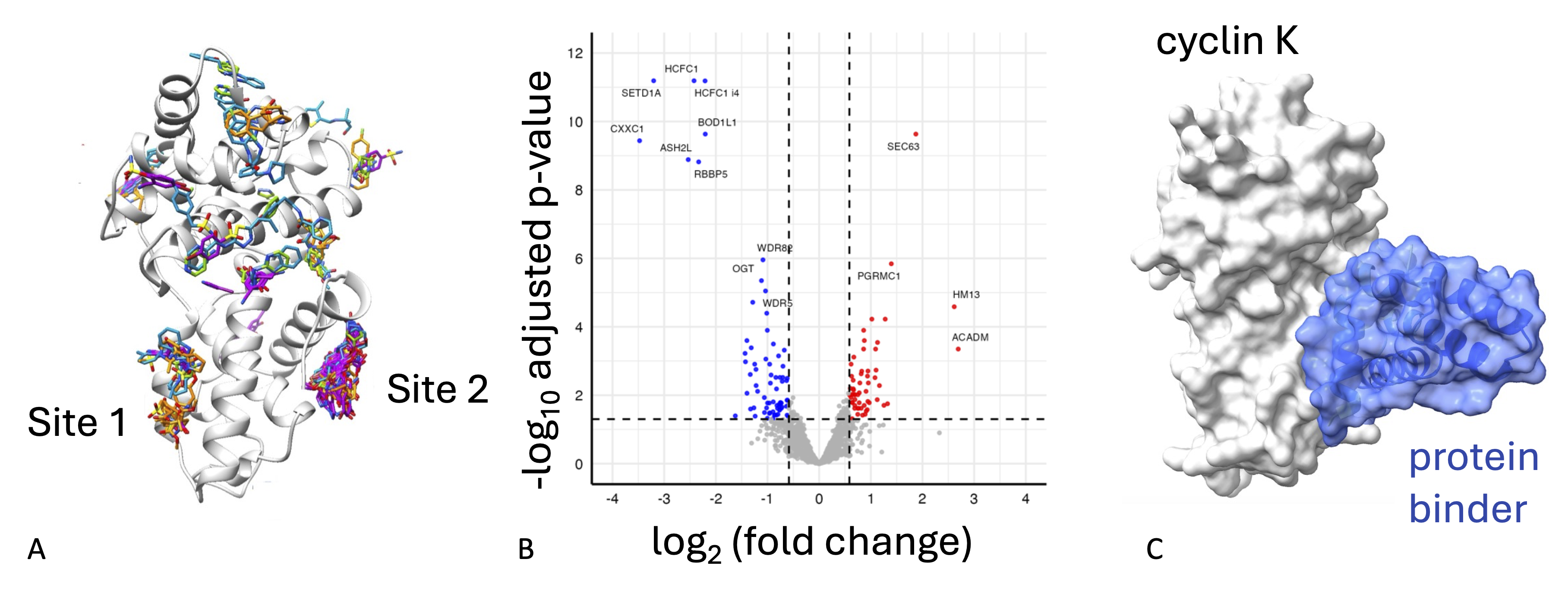
CDK12/13-cyclin K regulates transcription and promotes mRNA synthesis by RNA Polymerase II (RNA Pol II) through phosphorylation of the RNA Pol II C-terminal domain (RNA Pol II CTD). It also further facilitates productive elongation through phosphorylation of associated transcription factors and regulators. Early studies on CDK12 and cyclin K established that their depletion specifically impacts the expression of long-exon rich genes (>10 kb), notably DNA Damage Response genes (such as such as BRCA1, FANCF, and ERCC4). The direct interaction of CDK12-cyclin K with two large complexes, the PAF and COMPASS complexes contribute to the execution of its roles in transcription. The fragment map of cyclin K includes two rich and diverse fragment binding clusters, which collocate with cyclin K sites predicted from AlphaFold3 (AF3) modelling to bind to CDC73 and SETD1A (PAF1 and COMPASS complex subunits respectively). Using separation of function mutants (SOFs) combined with immunoprecipitation-mass spectrometry (IP-MS) we have identified cyclin K interactome perturbations consequent upon site-specific mutation. Using a pipeline that sequentially employs RFDiffusion, ProteinMPNN and AlphaFold2-Multimer we have designed site-selective small protein binders with nM affinity for cyclin K. Comparative mass spectrometry and functional analyses consequent to cyclin K SOF mutant or protein binder expression highlights the potential of protein binders as tools to probe protein function.
Highlights of our study
- FragLite mapping of cyclin K
- Identification of a SET1A binding hotspot required for recruitment of CDK12-cyclin K to the COMPASS complex
- Generation of a set of Separation of Function cyclin K mutants to further cyclin K mechanistic studies
- Design and characterisation of small protein binders that bind to cyclin K with nM affinity as functional probes.
PDB entries: 31TR, 31TO, 31UB, 31UA, 31TZ, 31TL, 31TK, 31TI, 31TC, 31TJ, 31TB, 31SX, 31TA, 31SZ, 31RZ, 31SE, 31SA, 31RY, 31RX, 31RV, 31MM, 31RW, 31RI, 31RN, 31RK, 31QC, 31QB, 31RQ, 31QA, 31QG, 31NZ, 31OA, 31NE, 31NJ, 31MO, 31MN, 31MI, 31NI, 31NG, 31NF, 31ND, 31NC, 31NB, 31NA, 31NH, 31MZ, 31NL, 31NM, 31NK
Collaborators: A Frey and M Troste (mass spectrometry), colleagues at the CRH Newcastle Drug Discovery Group and Newcastle University Structural Biology Facility
Associated publication: An integrated structural approach to identify tools to characterise the cyclin K interactome, (2026) Alshanbari et al manuscript in preparation