CDK2-cyclin A integration
A working kinase rarely acts alone. Cyclin-dependent kinase 2 (CDK2)-cyclin A is drawn into large assemblies that switch it on, turn it off, or mark its partners for destruction. Using cryo-electron microscopy (cryo-EM) we have caught two of these machines in the act, revealing how the same kinase surface is read out in completely different ways.
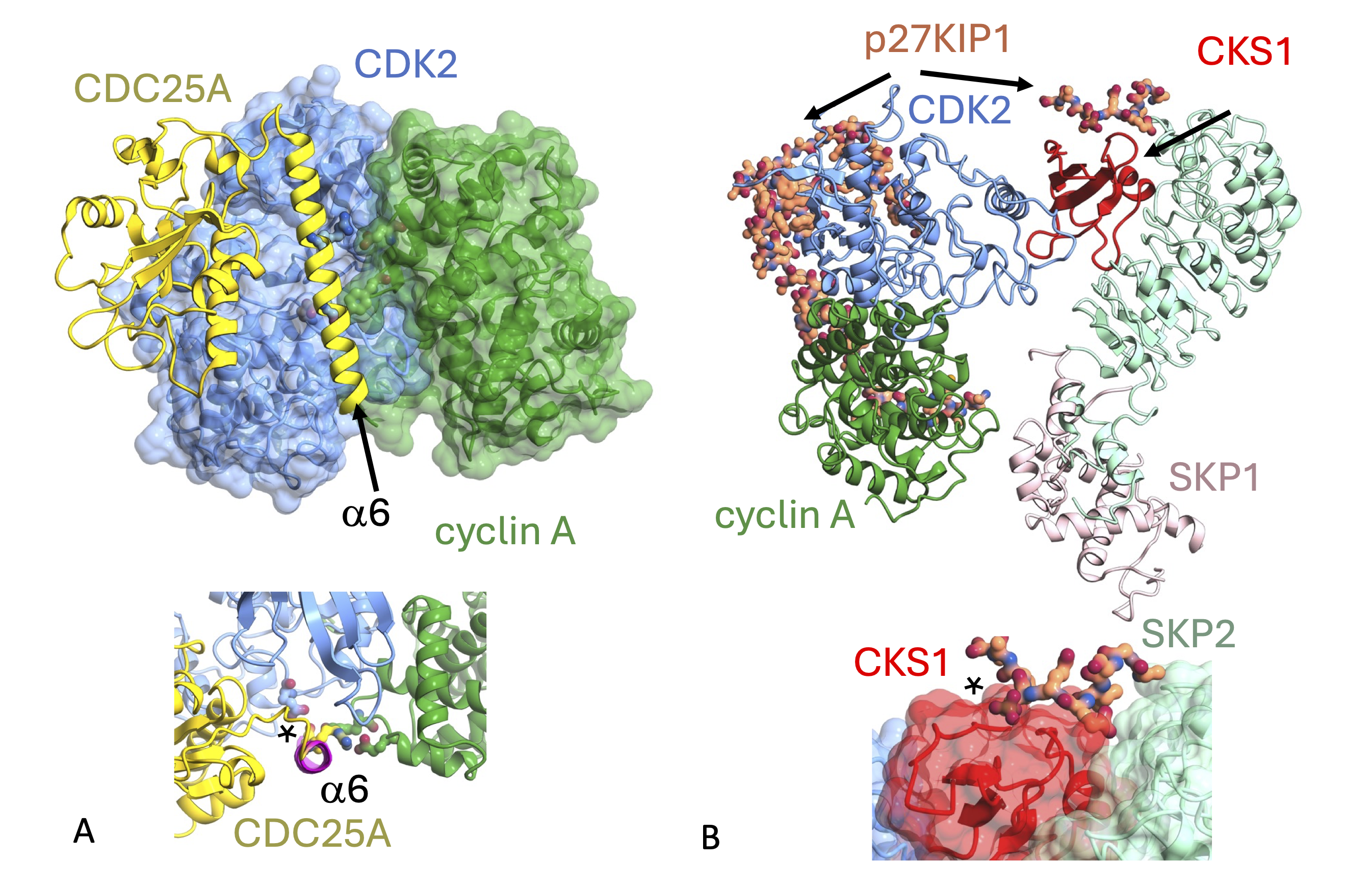
CDK2-cyclin A is regulated through protein-protein interactions (PPIs) that recruit the kinase module into many different complexes. To explore how PPIs regulate CDK2 phosphorylation status we have determined the structure of CDK2-cyclin A bound to the phosphatase CDC25A. To describe how CDK2-cyclin A-p27KIP1 is recognised by the E3 Ubiquitin ligase SCFSKP2 we have determined the structure of the hexameric CDK2-cyclin A-p27KIP1-SKP1-SKP2-CKS1 complex. Both structures were determined using the technique of cryo-electron microscopy (cryo-EM).
As a response to DNA damage the cell cycle arrests to provide time for the damage to be repaired. CDK2 drives the cell through the later stages of G1 phase (when bound to cyclin E) and through S-phase (when bound to cyclin A). To inhibit CDK2 activity in response to DNA damage, CDK2 is phosphorylated with the glycine-rich loop that forms part of the active site. When the damage is resolved, members of the CDC25 family of phosphatases dephosphorylate CDK2 to activate CDK2 activity. The structure of CDK2-cyclin A-CDC25A revealed how the phosphatase recognises its substrate at its active site and the critical importance of the CDC25A C-terminal helix (a6) for complex formation. A comparative structural analysis of CDK-containing complexes confirmed the functional importance of the conserved CDK1/2 GDSEID motif (single letter amino acid code). This motif is located on the CDK1/2 C-terminal lobe and is the binding site for multiple CDK2 regulators including CKS1 and KAP- the phosphatase that dephosphorylates Thr160 in the CDK2 activation segment. However, whilst the recognition sites for CDC25A and CKS1 partially overlap at the GDSEID region, the exact nature of their interactions are different and can be further fine-tuned by sequence differences in CDK1 and CDK2. Comparative sequence analysis and modelling studies using AlphaFold suggest that CDK4/6-cyclin D complexes are not CDC25 substrates supporting a model in which active site phosphorylation is not a mechanism used to inhibit CDK4/6 activity.
p27KIP1 is a member of the CIP/KIP family of CDK regulators that inhibit cell cycle CDKs. To promote cell cycle transitions p27KIP1 has to be degraded. To mark it for degradation, p27KIP1 bound to a CDK-cyclin is phosphorylated within its C-terminus by CDK1 or CDK2. This event signals p27KIP1 recruitment into the SCFSKP2 (S-phase kinase associated protein 1 (SKP1)-cullin-SKP2) complex, an E3 ubiquitin ligase that gates entry into the ubiquitin-dependent proteasome degradation pathway. Employing cryo-EM, we determined the structure of CDK2-cyclin A-p27KIP1 bound to SKP1-SKP2-CKS1. This hexameric structure supports previous analysis in which p27KIP1 was found to be structurally dynamic, transitioning from disordered to nascent secondary structure on target binding. We also studied the conformational space of the complex and uncovered a previously unidentified hinge motion giving rise to open and closed conformations. We proposed that that flexibility may contribute to p27KIP1 regulation by facilitating recognition with the larger SCFSKP2 complex.
Highlights of our study
- Determination of the structure of the CDK2-cyclin A-CDC25A complex
- Verification of the importance of the conserved GDSEID motif within the CDK2 C-terminal lobe as a sequence required for multiple CDK2-protein interactions
- Demonstration that the CDC25A C-terminal helix folds upon binding to CDK2-cyclin A and recognises the phosphorylation status of the CDK2 activation segment
- Comparative sequence analysis and AlphaFold modelling suggest CDK4/6-cyclin D complexes are not substrates for CDC25
- Determination of the structure of a hexameric CDK2-cyclin A-p27KIP1-SKP1-SKP2-CKS1 complex
Collaborators: James Ault, Jamie Blaza, (CDC25A complex) Michel Pagano (SKP2) and colleagues at the CRH Newcastle Drug Discovery Group and Newcastle University Structural Biology Facility.
PDB entries: 8ROZ (CDK2-cyclin A-CDC25A) and 8BYA (CDK2-cyclin A-p27KIP1-SKP1-SKP2-CKS1).
Electron Microscopy Data Bank entries: 19408 (CDK2-cyclin A-CDC25A) and 16325 (CDK2-cyclin A-p27KIP1-SKP1-SKP2-CKS1)
Associated publications:
Cryo-EM structure of SKP1-SKP2-CKS1 in complex with CDK2-cyclin A-p27KIP1, Rowland RJ et al., (2023) Sci Rep 13:10718. PMID: 37400515
Cryo-EM structure of the CDK2-cyclin A-CDC25A complex, Rowland RJ et al., (2024) Nat Commun 15:6807. PMID: 39122719