CDK1 structures
Every dividing cell depends on one enzyme above all others: cyclin-dependent kinase 1 (CDK1), which drives mitosis from dismantling the nucleus to splitting one cell into two. What makes it uniquely essential, and how does it differ from its near-twin CDK2 despite their near-identical active sites? The answer matters both for how the cell cycle is controlled and for designing inhibitors that can tell the two kinases apart.
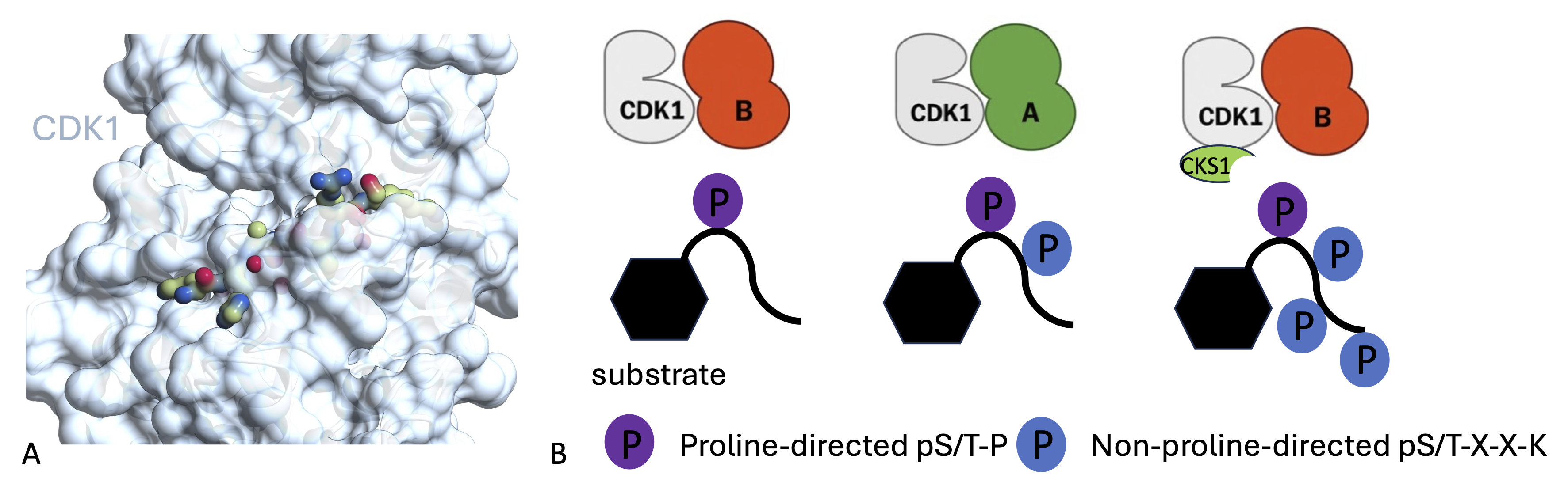
CDK1 is an essential member of the CDK family, and its sequence is highly conserved across eukaryotic species. It is required for successful completion of mitosis. CDK1 is activated by binding to its partners cyclin A or cyclin B and requires phosphorylation of Thr161 within the activation segment for full activity. During mitosis CDK1–cyclin B phosphorylates a large number of substrates to break down the nuclear membrane, separate the sister chromatids and drive the cell through cytokinesis. Together these events generate two daughter cells. Our determination of the crystal structures of CDK1–Cks1 and CDK1– cyclin B–Cks2 confirmed the conserved nature of the inactive monomeric CDK fold and its ability to be remodelled by cyclin binding. Relative to CDK2–cyclin A, CDK1–cyclin B is less thermally stable, has a smaller interfacial surface, is more susceptible to activation segment dephosphorylation and shows differences in the substrate sequence features that determine activity. In collaboration with the Ly group at the University of Dundee, we have shown how CDK1 activity and non-catalytic CDK1 subunits contribute to the choice of substrate and site of phosphorylation. A challenge for drug design is to identify compounds that selectively distinguish CDK1 and CDK2 as these CDKs share considerable sequence identity. CDK1 has a malleable fold that responds to cyclin and ATP-competitive inhibitor binding. We have used a range of biophysical techniques to compare the CDK1 and CDK2 folds in the cyclin-free and cyclin-bound states. These results hint at a different conformational energy landscape of cyclin-free CDK1 compared with cyclin-free CDK2, which is not apparent in cyclin-associated forms. This difference may underlie the essential cell-cycle role of CDK1. CDK1 can bind a sufficient set of cyclins to drive all phases of the cell cycle and can phosphorylate a broader range of substrates that includes sequences that do not conform to the canonical proline-directed substrate sequence.
Highlights of our study
- The first structure of CDK1 confirms the conserved nature of the inactive monomeric CDK fold and its ability to be remodelled by cyclin binding.
- Relative to CDK2–cyclin A, CDK1–cyclin B is less thermally stable, has a smaller interfacial surface and is more susceptible to activation segment dephosphorylation.
- Sequence conservation identifies potential sites of CDK1-protein interaction
- CDK1 and CDK2 exhibit differences in the substrate sequence features that determine activity both around the site of phosphotransfer and at +/- the RXL cyclin binding recruitment motif.
- CDK1 can phosphorylate an alternative S/T-X-X-K consensus sequence
- Demonstration that ATP-competitive inhibitors distinguish cyclin-free CDK1 from CDK2
- Determination of the first structures of ATP-competitive inhibitors bound to cyclin-free and cyclin-bound CDK1
Collaborators: Tony Ly (University of Dundee), Colleagues at the CRH Newcastle Drug Discovery Group and Newcastle University Structural Biology Facility.
PDB entries: 4YC6, 4YC3, 6GU3, 6GU2, 6GU4, 6GU6, 6GU7, 6GUB, 6GUC, 6GUE, 6GUF, 6GUH, 6GUK.
Associated publications:
CDK1 structures reveal conserved and unique features of the essential cell cycle CDK, (2015) Brown et al., Nature Communs 6:6769. PMID: 25864384
Differences in the conformational energy landscape of CDK1 and CDK2 suggest a mechanism for achieving selective CDK inhibition, (2019) Wood et al., Cell Chem Biol 26:121-130. PMID: 30472117
Cyclin A and Cks1 promote kinase consensus switching to non-proline-directed CDK1 phosphorylation, (2023) Al-Rawi et al., Cell Rep 42: 112139. PMID: 36840943