CRABP2–cyclin D3 binding
A protein can moonlight in an unexpected job. Cellular retinoic acid binding protein 2 (CRABP2), best known for ferrying vitamin-A signals into the nucleus, turns out to bind selectively to one cyclin, cyclin D3. We mapped where and how it binds, revealing a new docking site on the cyclin surface that could help integrate different signalling pathways.
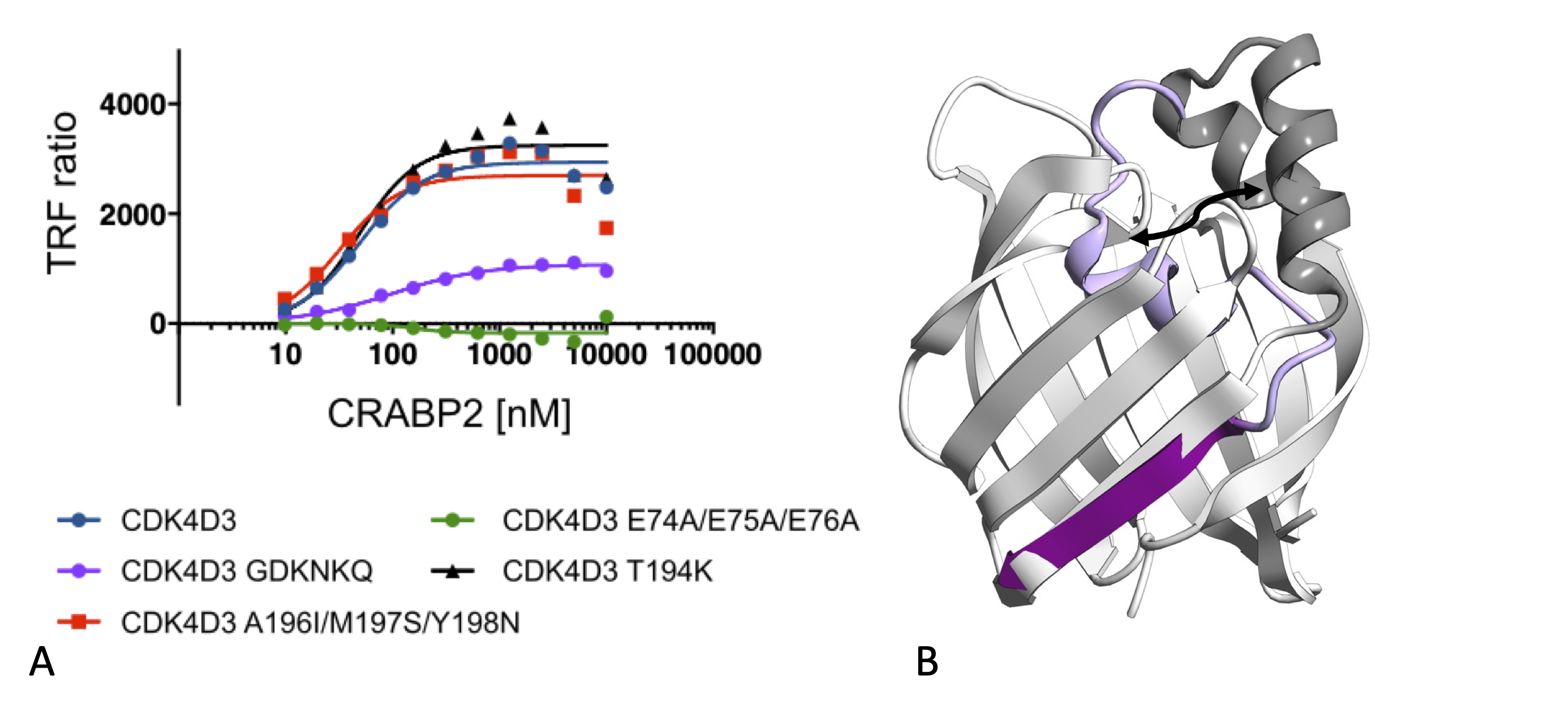
Cellular retinoic acid binding protein 2 (CRABP2) transports retinoic acid from the cytoplasm to the nucleus where it then transfers its cargo to retinoic acid receptor-containing complexes leading to activation of gene transcription. We demonstrate using purified proteins that CRABP2 is also a cyclin D3-specific binding protein and that the CRABP2 cyclin D3 binding site and the proposed CRABP2 nuclear localization sequence overlap. Both sequences are within the helix-loop-helix motif that forms a lid to the retinoic acid binding pocket. Mutations within this sequence (R30A/K31A) that block both cyclin D3 and retinoic acid binding promote formation of a CRABP2 structure in which the retinoic acid binding pocket is occupied by an alternative lid conformation (Figure panel B). Structural and functional analysis of CRABP2 and cyclin D3 mutants combined with AlphaFold models of the ternary CDK4/6-cyclin D3-CRABP2 complex supports the identification of an a-helical protein binding site on the cyclin D3 C-terminal cyclin box fold (Figure panel A). The model would suggest that like the cyclin recruitment site, this site is a hot-spot for cyclin D-protein interaction and can act to integrate signalling pathways to control CDK4/6-cyclin D function
Highlights of our study
- CRABP2 binds selectively to CDK4/6-cyclin D3 and not to CDK4/6 or CDK4/6-cyclin D1
- The CRABP2 nuclear localization sequence and cyclin D3 binding site overlap
- CRABP2 mutants adopt alternative monomeric CRABP2 conformations
- Cyclin D3 mutants identify a protein binding site on the C-terminal cyclin box fold
Collaborator: Ehmke Pohl and colleagues at the CRH Newcastle Drug Discovery Group and Newcastle University Structural Biology Facility
PDB entry: 7OXW, 7OXX
Associated publication: Structural requirements for the specific binding of CRABP2 to cyclin D3, (2024) Pastok et al Structure 32: 2301-2315. PMID: 39419021