Methods
Optogenetics:

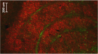
Fantastic method to dissect neuronal circuits into its components allowing for cell specific manipulations. Here the red area constitutes magno- and parvo cells in the thalamus. We managed to selectively target konio neurons in the green area.
Electrophysiology:
When you click on the movie above and have your loudspeaker on, you can here neurons at work, i.e. they “spike” in electrophysiological recordings. When we apply optogenetic green light stimulation, the neuron stops to be active. Electrophysiology is the gold standard to record neuronal activity directly with high temporal precision.
functional Magnetic Resonance Imaging (fMRI):
A great method to map functional brain organization at a mm-cm scale. Click on the movie to the left to see an experiment to reveal the retinotopic organization of visual cortex. Unfortunately the method measures neuronal activity only indirectly and with poor temporal resolution.
Pharmacology:

To generate the image to the left we superimposed an anatomical MRI scan visualizing a contrast agent with a functional image. The bright area in the MR image shows where we injected a drug to temporally shut down activity in the visual thalamus.
Selective Lesions:

Experimental aspiration lesions mimic conditions of brain injury and are used to induce and investigate mechanisms of brain reorganization. We use selective lesions of the visual cortex to study cortical blindness.